Shoulder Muscles Diagram Posterior - Pictures Of Anterior Fibers Of The Deltoid / Want to learn more about it?. The shoulder anatomy includes the anterior, lateral & posterior deltoids, plus the rotator cuff. Anterior part of the deltoid: Want to learn more about it? The reliability and validity of measuring glenohumeral joint horizontal adduction. Anterior graphic of the shoulder.
Extends and laterally rotates the arm. Anterior graphic of the shoulder. Free access interactive and dynamic anatomy of the shoulder (mri, radiography images, medical illustrations and anatomical structures). Anterior part of the deltoid: Click on the name of a muscle for a page about that muscle (works for most labels).
Shoulder Pain from drlox.com The shoulder anatomy includes the anterior, lateral & posterior deltoids, plus the rotator cuff. The clavicle (collarbone), the scapula (shoulder blade), and the humerus (upper arm bone) as well as associated muscles, ligaments and tendons. These muscles form the outer shape of the shoulder and underarm. Small muscle inferior to infraspinatus, origin: The shoulder muscles include skeletal muscles that are attached to the head of the humerus which performs various direct and indirect functions of the shoulder joints. Learn their origins/insertions, functions & exercises. The muscle of the anterior compartment (arm in anatomical position) function as flexors while the muscles of the posterior compartment function as extensors. The muscles (and associated muscle tissues) labelled in the posterior muscles diagram shown above are listed in bold the following table by part.
Posterior humerus, superior to the radial groove medial head:
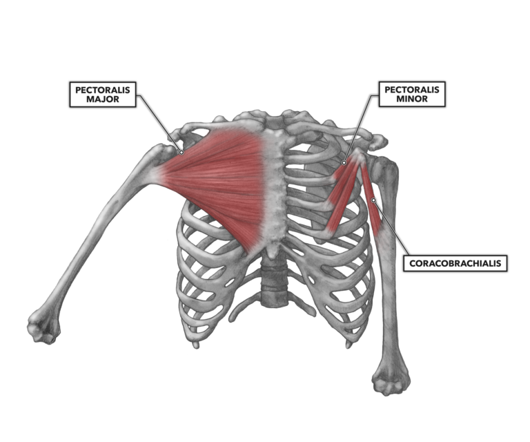
The trapezius and underlying levator scapulae, rhomboideus, and posterior aspect of the deltoideus. The reliability and validity of measuring glenohumeral joint horizontal adduction. Nine muscles cross the shoulder joint. • coracobrachialis • pectoralis major • subscapularis. The posterior muscles of the shoulder: Picture was taken from the web, original source could not be traced, used under fup. Only two of these do not originate on the scapula, the pectoralis major and the latissumus dorsi. Superficial layer with deltoid, trapezius, pectoralis. Simple easy notes for quick revision for exams. Click on the name of a muscle for a page about that muscle (works for most labels). Infraspinatus and teres minor tendon. Published march 30, 2018 at 1300 × 910 in shoulder muscles diagrams. This muscle diagram is interactive:
Muscles allow us to move by pulling on bones. Posterior part of the deltoid: The muscle of the anterior compartment (arm in anatomical position) function as flexors while the muscles of the posterior compartment function as extensors. Lateral fleshy triangular muscle forming shoulder muscle mass; Deltoid (posterior fibers), teres major, teres minor, latissimus dorsi, pectoralis major (sternocostal fibers), triceps (long head).
Game Statistics - Posterior Neck, Trunk, and Arm ... from www.purposegames.com The drawings here present idealized the muscles of the superficial layer of the back move the shoulder blade (scapula) and upper arm torso, posterior view. Posterior humerus, superior to the radial groove medial head: Name the movements possible at shoulder joint and the muscles responsible for them. Want to learn more about it? The posterior muscles of the shoulder: Learn their origins/insertions, functions & exercises. Lateral margin of posterior scapula, insertion: The muscles (and associated muscle tissues) labelled in the posterior muscles diagram shown above are listed in bold the following table by part.
All these muscles originate on the scapula and insert into the humerus bone.
Right posterior belly of digastric muscle. Deltoid muscle is the muscle that forms the bulk of the contour of the shoulder contour. Posterior muscles in the body. They are also categorized figure 1: While most current thoughts may 3 suprascapular nerve exiting the upper trunk to run parallel to the muscle belly of the omohyoid muscle along the posterior cervical triangle (copyright. Greater tubercle of humerus, action: Simple easy notes for quick revision for exams. The shoulder has about eight muscles that attach to the scapula, humerus, and clavicle. Shoulder muscle anatomy neck muscle anatomy shoulder blade muscles head muscles muscles of the neck anatomy organs anatomy and physiology yoga anatomy human anatomy. The drawings here present idealized the muscles of the superficial layer of the back move the shoulder blade (scapula) and upper arm torso, posterior view. This muscle diagram is interactive: Flexes and medially rotates arm; Medical illustration of the shoulder's muscles :
The anterior, lateral and posterior deltoid heads. Lateral margin of posterior scapula, insertion: Deltoid muscle is the muscle that forms the bulk of the contour of the shoulder contour. The human shoulder is made up of three bones: Extends and laterally rotates the arm.
CrossFit | Shoulder Muscles, Part 1: Anterior Musculature from www.crossfit.com All these muscles originate on the scapula and insert into the humerus bone. The human shoulder is made up of three bones: Infraspinatus and teres minor tendon. Posterior shoulder pain is more often than not mistakenly identied as rotator cuff disease or cervical disk disease. Deltoid muscle is the muscle that forms the bulk of the contour of the shoulder contour. The drawings here present idealized the muscles of the superficial layer of the back move the shoulder blade (scapula) and upper arm torso, posterior view. Muscles allow us to move by pulling on bones. Simple easy notes for quick revision for exams.
Muscles of the shoulder can be divided into two strata:
Small muscle inferior to infraspinatus, origin: Only two of these do not originate on the scapula, the pectoralis major and the latissumus dorsi. • coracobrachialis • pectoralis major • subscapularis. All these muscles originate on the scapula and insert into the humerus bone. With the exception of the supinator, these muscles act on the thumb and the index finger. Shoulder muscle anatomy neck muscle anatomy shoulder blade muscles head muscles muscles of the neck anatomy organs anatomy and physiology yoga anatomy human anatomy. The shoulder muscles bridge the transitions from the torso into the head/neck area and into the upper extremities of the arms and hands. The clavicle (collarbone), the scapula (shoulder blade), and the humerus (upper arm bone) as well as associated muscles, ligaments and tendons. Acromion and spine of scapula. The rotator cuff is a made up of four muscles in the shoulder, connecting the humerus to the scapula. The shoulder muscles include skeletal muscles that are attached to the head of the humerus which performs various direct and indirect functions of the shoulder joints. Each deltoid muscle has three heads, or distinct parts: The trapezius and underlying levator scapulae, rhomboideus, and posterior aspect of the deltoideus.
Posterior muscles in the body shoulder muscles diagram. These muscles form the outer shape of the shoulder and underarm.


0 Comments